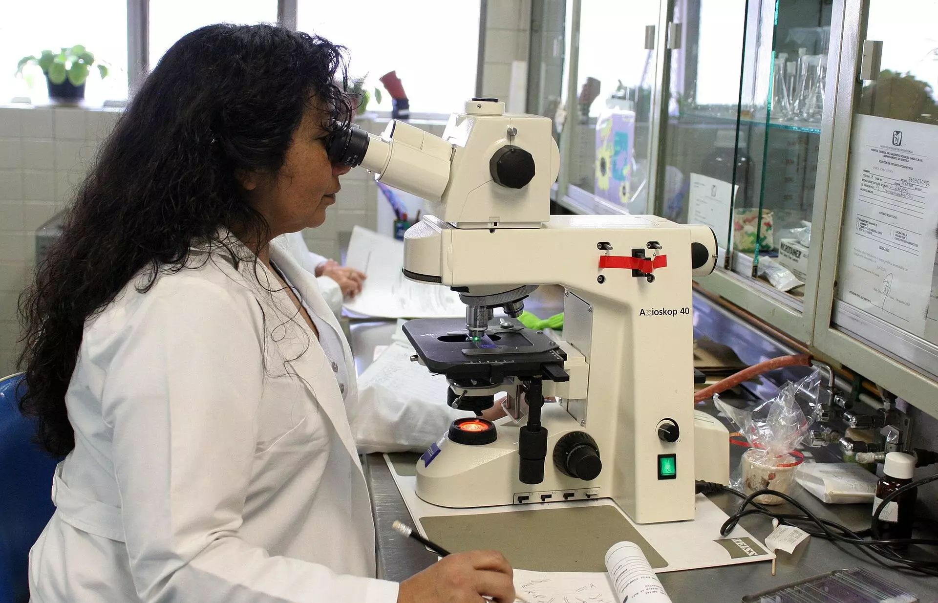
When utilizing a microscope to view biological samples, it is essential to consider the disturbance caused by the different mediums present. The light beam traveling through the lens of the objective can be altered if it encounters a medium distinct from the sample. For instance, if a watery sample is observed with a lens surrounded by air, the light rays will bend more sharply in the air compared to the water. This phenomenon results in the measured depth within the sample appearing smaller than its actual depth, leading to a flattened appearance.
Historically, theories dating back to the 80s have aimed to provide a corrective factor for determining the depth distortion in microscopy. However, these theories assumed a constant corrective factor, irrespective of the sample’s depth. It was not until the 90s when Nobel laureate Stefan Hell highlighted the possibility of depth-dependent scaling. Associate Professor Jacob Hoogenboom of Delft University of Technology notes the gap in addressing this issue over the years.
Sergey Loginov, a former postdoc at Delft University of Technology, has conducted calculations and developed a mathematical model to demonstrate that samples exhibit stronger flattening closer to the lens and less farther away. Subsequent laboratory experiments by Ph.D. candidate Daan Boltje and postdoc Ernest van der Wee have confirmed the depth-dependent nature of the corrective factor. Their findings have been published in the journal Optica.
Ernest Van der Wee emphasizes the significance of their research by providing a web tool and software that allow researchers to determine the precise corrective factor for their experiments. By utilizing these tools, researchers can accurately extract protein structures from biological systems using electron microscopy. This enhanced precision saves time and resources by avoiding samples that do not align with the biological target, enabling the study of more pertinent proteins and biological structures.
Daan Boltje underscores the importance of precise depth determination in studying proteins within biological systems. Understanding the exact structure of proteins is vital for deciphering abnormalities and diseases, ultimately paving the way for effective treatments. By implementing depth-dependent correction in microscopy, researchers can streamline their investigations and focus on relevant biological components.
The web tool provided allows researchers to input various experiment details, such as refractive indices, aperture angle, and light wavelength. The tool generates a curve depicting the depth-dependent scaling factor, which can be exported for individual use. Furthermore, researchers can compare the results with existing theories to gain a comprehensive understanding of the corrective process in microscopy.


Leave a Reply