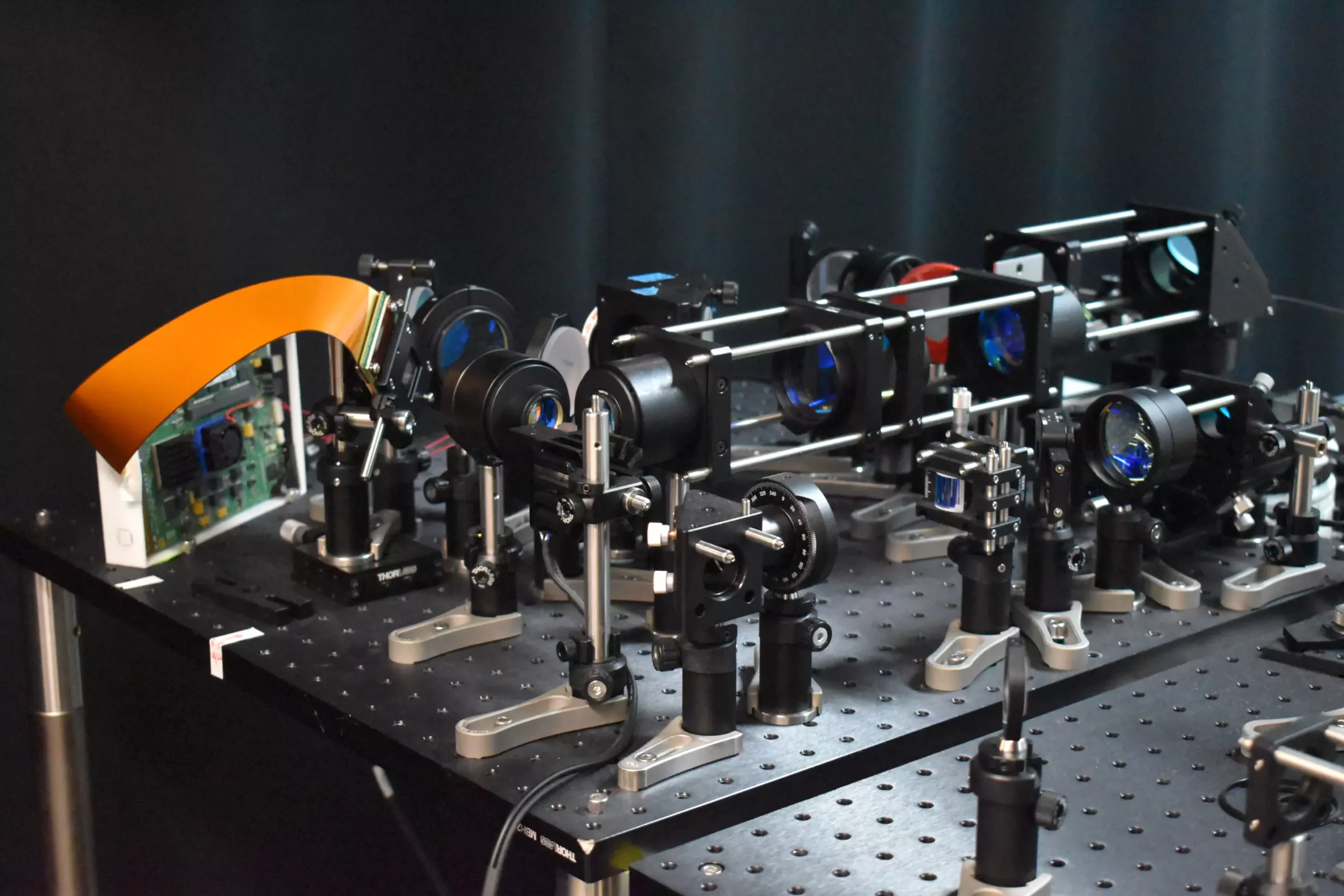
The development of a new two-photon fluorescence microscope is set to revolutionize the field of neuroscience by capturing high-speed images of neural activity with cellular resolution. This breakthrough technology allows researchers to image neural networks in real time, providing valuable insights into essential brain functions such as learning, memory, and decision-making.
Unlike traditional two-photon microscopy methods, this innovative microscope offers faster imaging capabilities with reduced harm to brain tissue. By replacing point illumination with line illumination, the new approach enables in vivo imaging of neuronal activity in a mouse cortex at speeds ten times faster while decreasing the laser power on the brain by more than tenfold. This advancement allows for a clearer view of how neurons communicate and interact in real-time, paving the way for a better understanding of brain function and the pathology of neurological diseases.
The key to this groundbreaking technology lies in the adaptive sampling strategy employed by researchers. Instead of using a single point of light to image specific areas of the brain, a short line of light is utilized to illuminate active neurons. This innovative approach enables a larger area to be excited and imaged simultaneously, significantly speeding up the imaging process. By targeting only the neurons of interest and not the background or inactive areas, the total light energy deposited on the brain tissue is reduced, minimizing the risk of potential damage.
Utilizing Digital Micromirror Device (DMD)
Researchers have leveraged a digital micromirror device (DMD), containing thousands of tiny mirrors that can be individually controlled, to shape and steer the light beam. This precise targeting of active neurons is achieved by turning individual DMD pixels on and off, adjusting to the neuronal structure of the brain tissue being imaged. The DMD also mimics high-resolution point scanning, enabling the reconstruction of high-resolution images from fast scans. This critical advancement allows for quick identification of neuronal regions of interest, essential for subsequent high-speed imaging and adaptive sampling.
Uncovering Rapid Neural Events
The new microscope was demonstrated by imaging calcium signals, indicators of neural activity, in living mouse brain tissue at a speed of 198 Hz. This remarkable speed surpasses traditional two-photon microscopes, capturing rapid neuronal events that would be missed by slower imaging methods. The adaptive line-excitation technique, combined with advanced computational algorithms, enables the isolation of individual neuron activity. This capability is vital for the accurate interpretation of complex neural interactions and understanding the functional architecture of the brain.
Future Directions
Moving forward, researchers are focused on integrating voltage imaging capabilities into the microscope to provide a direct and rapid readout of neural activity. Additionally, they plan to apply the new method to real neuroscience applications, such as observing neural activity during learning and studying brain activity in disease states. Efforts are also underway to enhance the microscope’s user-friendliness and reduce its size to expand its utility in neuroscience research.
This innovative two-photon fluorescence microscope represents a significant advancement in real-time imaging of neural activity, offering a powerful tool for studying dynamic neural processes with minimal risk to living tissue. By pushing the boundaries of traditional microscopy techniques, researchers are poised to gain unprecedented insights into brain function, paving the way for improved treatments for neurological diseases and a deeper understanding of the complexities of the human brain.


Leave a Reply