
In a groundbreaking study published in the journal Optica, researchers at HHMI’s Janelia Research Campus have revolutionized the field of microscopy by adapting techniques from astronomy to enhance image clarity. This innovative approach presents a faster and more cost-effective method for biologists to obtain high-resolution microscopy images, ultimately advancing the capabilities of biological research.
Traditionally, astronomers have utilized sophisticated methods to correct the distortion of light caused by atmospheric conditions, thereby improving the quality of images captured by telescopes. Building upon this foundation, microscopists have endeavored to apply similar principles to biological samples, which also exhibit light bending and distortions. However, current adaptive optics methods are intricate, expensive, and time-consuming, limiting their accessibility to many research laboratories.
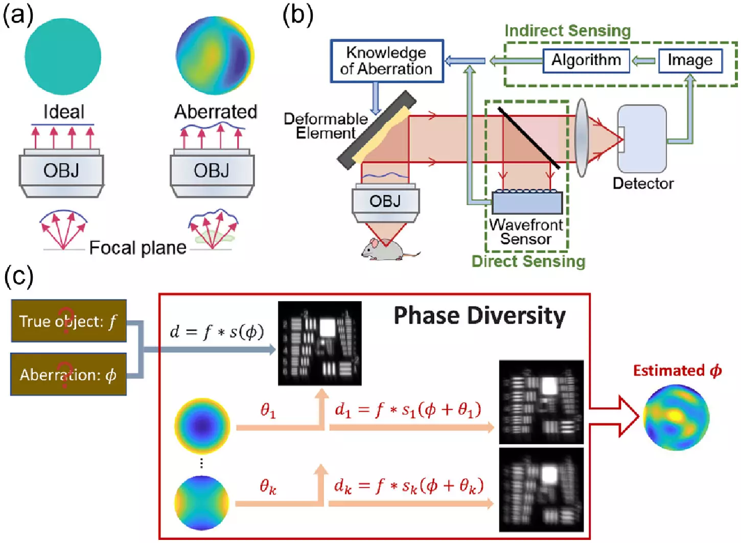
To address these challenges, the research team at Janelia Research Campus turned to a novel approach known as phase diversity. While commonly used in astronomy, phase diversity techniques had not yet been explored in the life sciences. By integrating additional images with known aberrations into the original blurry image, the phase diversity method offers a unique solution to unblurring distorted images without requiring extensive modifications to existing microscopy systems.
To demonstrate the effectiveness of the new method, the team first adapted the astronomy algorithm for microscopy applications and conducted rigorous simulations to validate its performance. Subsequently, they designed a specialized microscope equipped with a deformable mirror and additional lenses to introduce known aberrations. These minor modifications, coupled with advancements in software development, enabled the researchers to significantly enhance image clarity and resolution.
In a series of experiments, the team successfully calibrated the deformable mirror of the microscope at a rate that was 100 times faster than traditional methods. Furthermore, they demonstrated the capability of the new technique to detect and correct randomly generated aberrations, resulting in clearer images of fluorescent beads and fixed cells. This notable advancement paves the way for future testing on live cells and tissues, as well as the integration of phase diversity into more complex microscopy systems.
The innovative approach introduced by the researchers offers a faster and more cost-effective alternative to existing adaptive optics techniques. By simplifying the implementation process and streamlining image correction procedures, the new method has the potential to democratize the use of adaptive optics in biological research. By facilitating clearer visualization of intricate biological structures, this technology promises to expand the horizons of scientific exploration and discovery.
Leave a Reply