
The advancement of microscopy techniques has revolutionized the field of biological research, allowing scientists to explore the intricate world of viruses, proteins, and molecules at a cellular level. However, traditional microscopy methods often come with limitations in terms of resolution and sample preparation. Researchers at the University of Tokyo have recently made a groundbreaking development in mid-infrared microscopy, significantly improving the resolution and allowing for the visualization of structures inside living bacteria at the nanometer scale.
Super-resolution fluorescent microscopes, while powerful, require the labeling of specimens with fluorescence, which can be toxic and lead to sample damage. Additionally, extended light exposure in fluorescent microscopy can bleach samples, rendering them unusable for further analysis. On the other hand, electron microscopes provide impressive detail, but the requirement for samples to be placed in a vacuum makes it impossible to study live samples. Mid-infrared microscopy offers a solution by providing both chemical and structural information about live cells without the need for labeling or damaging the samples. However, its resolution capabilities have historically been limited compared to other microscopy techniques.
The team at the University of Tokyo has successfully addressed the resolution limitations of traditional mid-infrared microscopy by achieving a spatial resolution of 120 nanometers, a significant improvement from the typical 3-micron resolution. This groundbreaking development, published in Nature Photonics, marks a 30-fold enhancement in resolution, enabling researchers to observe intracellular structures with unprecedented clarity. By utilizing a “synthetic aperture” technique that combines multiple images taken from different illuminated angles, the researchers were able to create a clearer overall picture of the samples.
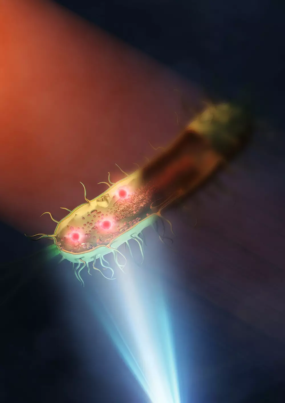
To overcome the limitations of conventional mid-infrared microscopy, the researchers implemented innovative strategies in their experimental setup. By placing the sample, bacteria such as E. coli and Rhodococcus jostii RHA1, on a silicon plate that reflected visible light and transmitted infrared light, they were able to eliminate the need for dual lenses that absorbed mid-infrared light. This approach allowed for better illumination of the samples with mid-infrared light, resulting in more detailed and high-resolution images of the intracellular structures of bacteria.
The enhanced resolution capabilities of the improved mid-infrared microscope have far-reaching implications for various fields of research, including infectious diseases and antimicrobial resistance. The ability to visualize and study intracellular structures at the nanometer scale opens up new opportunities for investigating biological processes and developing more accurate imaging techniques in the future. With further advancements in lens technology and the use of shorter wavelengths of visible light, the spatial resolution of mid-infrared microscopy could potentially reach below 100 nanometers, unlocking even greater potential for biological research.
The breakthrough achieved by the University of Tokyo researchers in improving mid-infrared microscopy represents a significant advancement in the field of biological imaging. By overcoming the resolution limitations of traditional techniques, this innovative approach opens up new possibilities for studying live cells and understanding complex biological mechanisms at a level of detail never before possible. The continuous refinement of mid-infrared microscopy holds promise for further discoveries and advancements in biological research, paving the way for new insights into the microscopic world of bacteria and beyond.
Leave a Reply